Schilling, Kurt G.; Ramadass, Karthik; Sairanen, Viljami; Kim, Michael E.; Rheault, Francois; Newlin, Nancy; Nguyen, Tin; Barquero, Laura; D’archangel, Micah; Gao, Chenyu; Topolnjak, Ema; Khairi, Nazirah Mohd; Archer, Derek; Beason-Held, Lori L.; Resnick, Susan M.; Hohman, Timothy; Cutting, Laurie; Schneider, Julie; Barnes, Lisa L.; Bennett, David A.; Arfanakis, Konstantinos; Vinci-Booher, Sophia; Albert, Marilyn; Moyer, Daniel; Landman, Bennett A. “.” Human brain mapping, vol. 46, no. 3, 2025, e70143, .Ěý
Head movement during MRI scans can lead to errors that affect the accuracy of results. However, no comprehensive study has been done to fully understand how motion impacts scans across different groups of people and types of studies. To address this, we conducted a large-scale study with three goals. First, we aimed to examine how much people move during MRI scans across 13 different groups, which included nearly 17,000 scan sessions from people of various ages (ranging from babies to elderly adults) and conditions (such as cognitive impairments and developmental delays). Second, we wanted to see if the latest image processing techniques could reduce errors caused by movement, especially when comparing scans taken at different times from the same person. Lastly, we looked at whether there were differences in brain connections between people who moved a lot during the scans and those who didn’t. Our findings showed that, on average, people move about 1–2 millimeters per minute, mostly in the front-to-back direction or by rotating their head from side to side. We also found that modern image processing techniques are effective at reducing the effects of motion, so any bias in the results was not noticeable with current methods. Finally, there were no significant differences in brain structure or connectivity between people who moved a lot and those who stayed still. In conclusion, understanding how motion affects scans and improving the way we process images can help make MRI results more accurate, even when there is head movement.Ěý
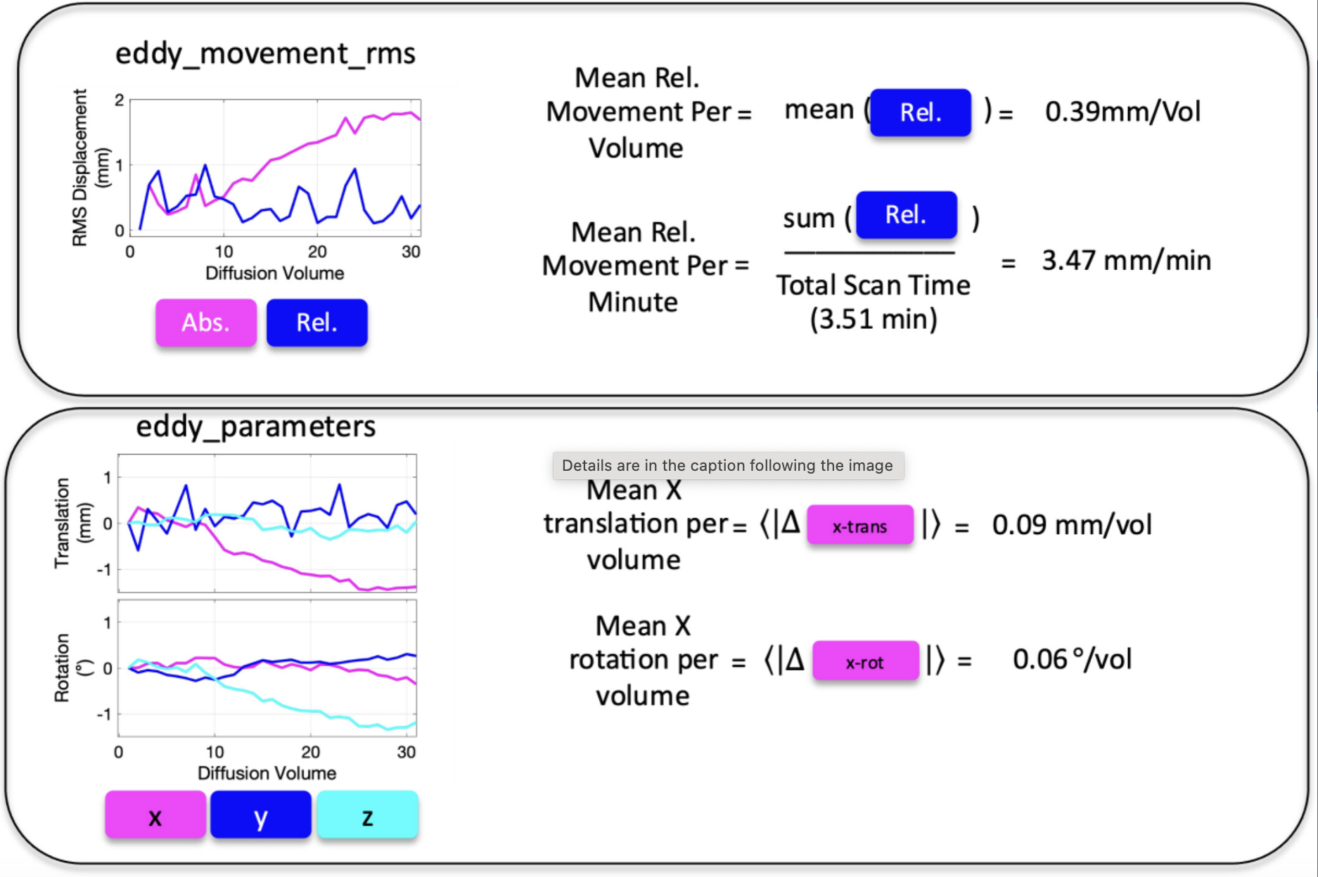
FIGURE 1Ěý
Derivation of head motion descriptors. All motion parameters were derived from output of FSL software’sĚýeddyalgorithm: Eddy_movement_rms (top) and eddy_parameters (bottom) files. From eddy_movement_rms, the “mean relative movement per imaging volume” (units of mm/volume) was calculated as the mean of the relative RMS displacement. This measure can be normalized by total scan time to the “mean relative movement per minute” (units of mm/min). From eddy_parameters, measures of absolute translation and rotation are given with respect to the first imaging volume. To calculate the “mean translation per volume”, we take the mean of the absolute values of differences from volume to volume (units of mm/vol). This is repeated for translation alongĚýx,Ěýy, andĚýzĚýaxes, as well as rotation around L/R, P/A, and I/S axis (units of degrees/vol).Ěý